
Using bio-based CaCO3 functionalized sediment to simultaneously remove algae and COD through adsorption and sedimentation in water source reservoirs
Supplementary Information
a School of Civil Engineering, Chang’an University, Xi’an 710064, China.
b Key Laboratory of Environmental Aquatic Chemistry, State Key Laboratory of Regional Environment and Sustainability, Research Center for Eco-Environmental Sciences, Chinese Academy of Sciences, Beijing 100085, China.
c University of Chinese Academy of Sciences, Beijing 100049, China.
* Corresponding to: Jinyi Qin (jinyi.qin@chd.edu.cn), Ming Su (mingsu@rcees.ac.cn)
Figures and/or tables are provided below as the supplementary evidences to the main text.
Supplementary methods
Langmuir equation (Veith and Sposito, 1977)
The equation for the pseudo-first-order kinetic model is (Eq. S1 and S2):
\(\ln(q_{e,cal} - q_{t}) = \ln q_{e} - k_{1}t\) (S1)
\(\ln(q_{e} - q_{t}) = \ln q_{e} - k_{1}t\) (S2)
The equation for the pseudo-second-order kinetic model is (Eq. S3):
\(\frac{t}{q_{t}} = \frac{1}{k_{2}q_{e}^{2}} + \frac{t}{q_{e}}\) (S3)
\(q_{e} = \frac{C_{i} - C_{f}}{S}\) (S4)
\(q_{t} = \frac{C_{i} - C_{t}}{S}\) (S5)
Where, \(q_{e}\) (μg g−1) (Almomani and Bhosale, 2021) is the experimental equilibrium adsorption capacity of the adsorbent; \(q_{t}\) (μg g−1) (Almomani and Bhosale, 2021) is the equilibrium adsorption amount of the adsorbent at time \(t\) (min); \(q_{e,cal}\) and \(q_{e}\) (μg g−1) are the theoretical and actual equilibrium adsorption amounts, respectively; \(k_{1}\) (min−1) is the pseudo-first-order kinetic model constant, \(k_{2}\) (g (mg min) −1 ) is the pseudo-second-order kinetic model constant.
The equilibrium adsorption data were analyzed using both Langmuir (Eq. S6) and Freundlich (Eq. S8) isotherm models (Almomani and Bhosale, 2021) to characterize the adsorption process.
\(q_{e} = \frac{q_{m}K_{L}C_{e}}{1 + K_{L}C_{e}}\) (S6)
\(R_{L} = \frac{1}{1 + K_{L}C_{e}}\) (S7)
\(q_{e} = K_{F}\left( C_{e} \right)^{\frac{1}{n}}\) (S8)
Where, \(q_{m}\) is the maximum Chl-a concentration adsorbed on unit mass of micro-nano CaCO3 (μg g−1), \(K_{L}\)is the Langmuir adsorption constant (μg L−1); \(R_{L}\)is the separation coefficient (Eq. S6); \(K_{F}\) is the Freundlich adsorption constant (μg g−1) (L μg−1)1/n; \(C_{e}\) is the adsorption equilibrium solution concentration (μg L-1), and \(1/n\) is the adsorption intensity coefficient (Huang et al., 2020). A value of \(1/n\) in the range \(0 < 1/n < 1\) indicates favorable adsorption with high affinity, while \(1/n > 1\) suggests poor adsorption and low surface affinity. The closer \(1/n\) is to 0, the stronger the adsorption intensity; conversely, larger \(1/n\) values reflect increased surface heterogeneity and weaker adsorption.
A dimensionless separation factor (\(R_{L}\)) is introduced from the Langmuir equation to evaluate whether the adsorption of bio-CaCO3 to algae is a favorable spontaneous reaction (Eq. S9).
\(R_{L} = \frac{1}{(1 + K_{L}C_{0})}\) (S9)
When \(R_{L} > 1\), it is unfavorable for the material’s adsorption; when \(R_{L} = 1\), the material’s adsorption is linear; when \(R_{L}\)=0, the material’s adsorption is irreversible; when \({0 < R}_{L} < 1\), it indicates that the material’s adsorption process is favorable. In the range of 0~1, the larger the value, the more favorable it is for the material to remove pollutants.
Young’s equation (Novak and Brune, 1985)
\[ \begin{aligned} G_{h_{0}}^{AB} &= 2\Bigg[\sqrt{\gamma_{\omega}^{+}}\left(\sqrt{\gamma_{f}^{-}} + \sqrt{\gamma_{m}^{-}} - \sqrt{\gamma_{\omega}^{-}}\right) \\ &\quad + \sqrt{\gamma_{\omega}^{-}}\left(\sqrt{\gamma_{f}^{+}} + \sqrt{\gamma_{m}^{+}} - \sqrt{\gamma_{\omega}^{+}}\right) \\ &\quad - \sqrt{\gamma_{f}^{-}\gamma_{m}^{+}} - \sqrt{\gamma_{f}^{+}\gamma_{m}^{-}}\Bigg] \\[1em] G_{h_{0}}^{LW} &= -2\left(\sqrt{\gamma_{m}^{LW}} + \sqrt{\gamma_{\omega}^{LW}}\right)\left(\sqrt{\gamma_{f}^{LW}} + \sqrt{\gamma_{\omega}^{LW}}\right) \end{aligned} \] (S10)
\[ \Delta G_{h_{0}}^{EL} = \frac{\varepsilon_{r}\varepsilon_{0}\kappa}{2}(\xi_{m}^{2} + \xi_{f}^{2})\lbrack(1 - {\cot h}(\kappa h_{0}) + \frac{2\xi_{m}\xi_{f}}{\xi_{m}^{2} + \xi_{f}^{2}}(\csc h(\kappa h_{0}))\rbrack \] (S11)
Where \(G_{h_{0}}^{AB}\), \(G_{h_{0}}^{LW}\) and \(\Delta G_{h_{0}}^{EL}\) is the individual interaction per unit area at \(h_0\), \(\gamma^-\) and \(\gamma^+\) are the electron donor and electron acceptor components of surface tension, respectively, and \(\gamma^{LW}\) is the Lifshitz–van der Waals component. The subscript \(w\) denotes the aqueous medium, and \(\theta\) is the contact angle \(\zeta m\) and \(\zeta p\) (mV) are the membrane and particle zeta potentials.
\[ \begin{aligned} \frac{(1 + \cos\varphi)}{2}\gamma_{l}^{\mathrm{Tol}} &= \sqrt{\gamma_{l}^{\mathrm{LW}}}\sqrt{\gamma_{s}^{\mathrm{LW}}} + \sqrt{\gamma_{l}^{-}}\sqrt{\gamma_{s}^{+}} + \sqrt{\gamma_{l}^{+}}\sqrt{\gamma_{s}^{-}} \\ \gamma^{\mathrm{Tol}} &= \gamma^{\mathrm{LW}} + \gamma^{\mathrm{AB}} \end{aligned} \] (S12)
\(\gamma^{AB} = 2\sqrt{\gamma_{l}^{-}\gamma_{s}^{+}}\) (S13)
\(\kappa = \sqrt{\frac{e^{2}\sum_{}^{}{n_{i}z_{i}}}{\varepsilon\varepsilon_{0}\kappa T}}\) (S14)
Where \(e\) is the elementary charge, \(i\) is the number concentration of the ions in the dispersed solution, \(Z_{i}\) is the valence of the ions, \(k\) is the Boltzmann constant, and \(T\) is the absolute temperature, (\(\varepsilon_{0}=8.85\times 10^{-12}\text{CV}^{-1}m^{-1}\)) (Oss, 1993) is the dielectric constant of vacuum, (\(\varepsilon = 78.5\)) is the dielectric constant of water.
DFT Calculations
All density functional theory (DFT) calculations were performed using the Vienna Ab initio Simulation Package (VASP) (Kresse and Furthmüller, 1996). The exchange–correlation interactions were treated using the generalized gradient approximation (GGA) with the Perdew–Burke–Ernzerhof (PBE) functional (Perdew et al., 1996). To account for dispersion forces, Grimme’s DFT-D3 empirical correction was applied (Grimme et al., 2010). A vacuum slab of approximately 15 Å was introduced perpendicular to the surface to eliminate interactions between periodic images.
A plane-wave cutoff energy of 450 eV was used. Brillouin zone sampling was conducted using a Γ-centered Monkhorst–Pack k-point mesh of 3 × 3 × 1 (Monkhorst and Pack, 1976). Full geometric optimizations were carried out until the residual force on each atom was less than 0.02 eV/Å and the total energy convergence threshold was set to 10-5 eV.
The adsorption energy (\(\Delta E\)) was calculated according to Eq. S15:
\(\Delta E = E_{ads/surf} - E_{\text{surf}} - E_{\text{ads}}\) (S15)
where \(E_{\text{ads/surf}}\) is the total energy of the adsorbate–substrate system, \(E_{surf}\) is the energy of the clean surface, and \(E_{ads}\) is the energy of the isolated adsorbate in vacuum.
Response surface design (Anderson-Cook et al., 2009)
A Box Behnken Design (BBD) was employed to optimize the algae removal process with minimal experimental runs. Three key factors—mass fraction of nano-CaCO3 (A), the molar ratio of Ca2+ to CO32- (B), and reaction time (C)—were selected as independent variables. The removal efficiencies of Chl-a (\(Y_1\)) and COD (\(Y_2\)) served as response variables. A total of 17 experimental runs were determined (S16). The independent variables were coded as \(X_1\), \(X_2\) and \(X_3\) (S17), and a second-order polynomial model was used to fit the response data.
\(N=2^k+2k+C_0\) (S16)
Where \(N\) is the number of experiments; \(k\) is the number of experimental factors,\(k = 3\); \(C_{0}\) is the number of repeated experiments at the center point, \(C_{0} = 3\).
\(Z_i=\frac{X_i-X_0}{\Delta X}\) (S17)
Where: \(Z_{i}\) is the dimensionless coded value of the i-th influencing factor; is the actual value of the i-th influencing factor; \(X_{0}\) is the actual value of \(X_{i}\ \)at the center point; \(\mathrm{\Delta}X\) is the difference between the actual value of the high level and the actual value of the center level of each influencing factor.
The quadratic polynomial model (S18) was applied to predict system behavior and identify optimal conditions:
\(Y = \beta_{0} + \sum_{i = 1}^{K}\beta_{i}X_{i} + \sum_{i = 1}^{k - 1}{\sum_{j = i + 1}^{k}{\beta_{ij}X_{i}}}X_{j} + \sum_{i = 1}^{k}{\beta_{ij}X_{i}^{2}}\) (S18)
where: \(Y\) represents the predicted response (removal efficiency), \(\beta_{0}\) is the constant coefficient, \(\beta_{i}\) are linear effect coefficients, \(\beta_{ij}\) are interaction effect coefficients, \(\beta_{ii}\) are quadratic effect coefficients, \(X_{i}\) and \(X_{j}\) denote coded values of independent variables.
Supplementary figures
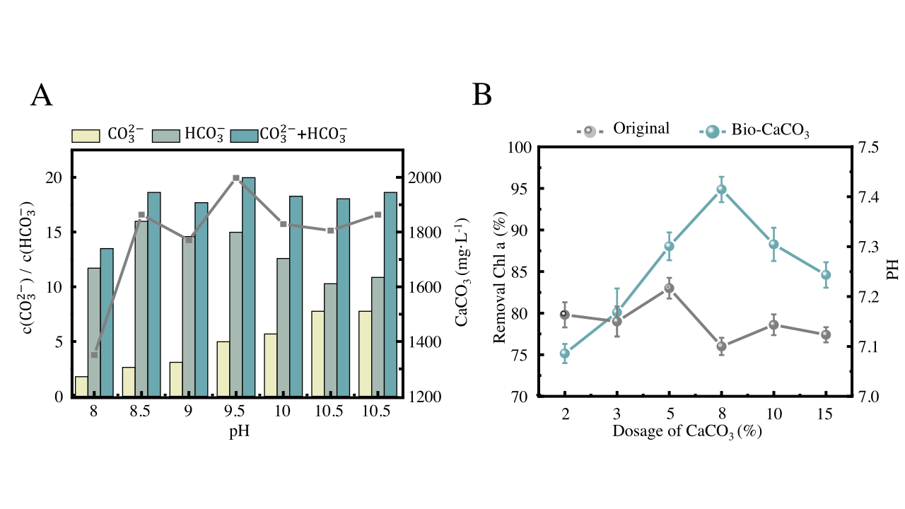
Supplementary Fig. 1
Supplementary Fig. 2
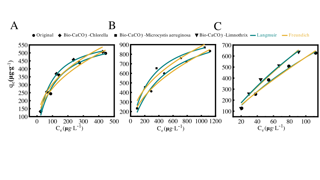
Supplementary Fig. 3
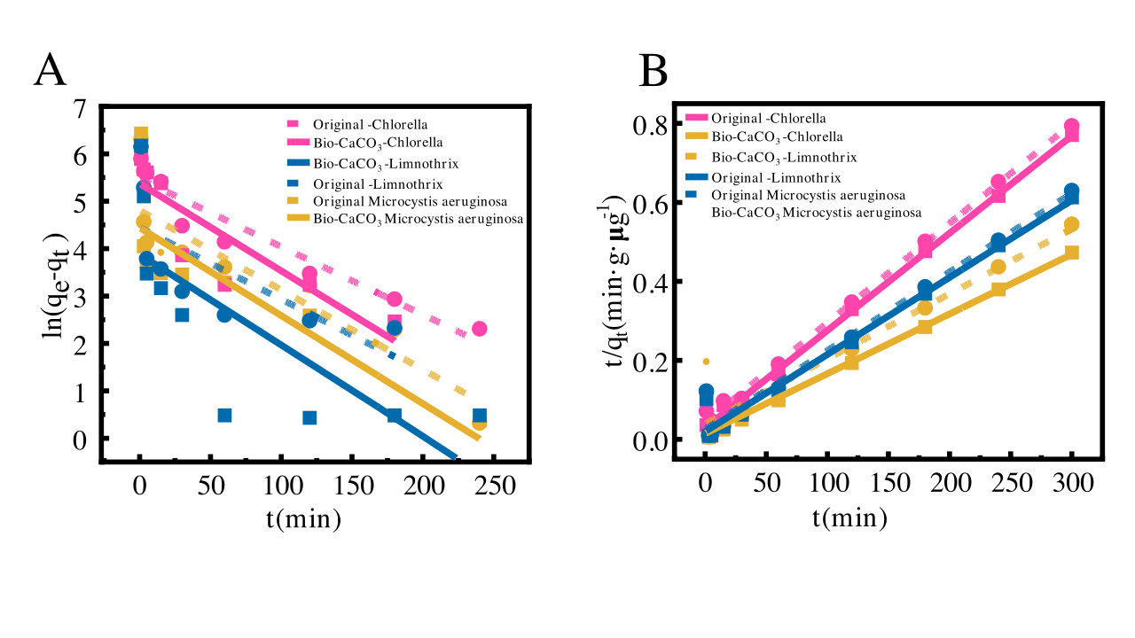
Supplementary Fig. 4
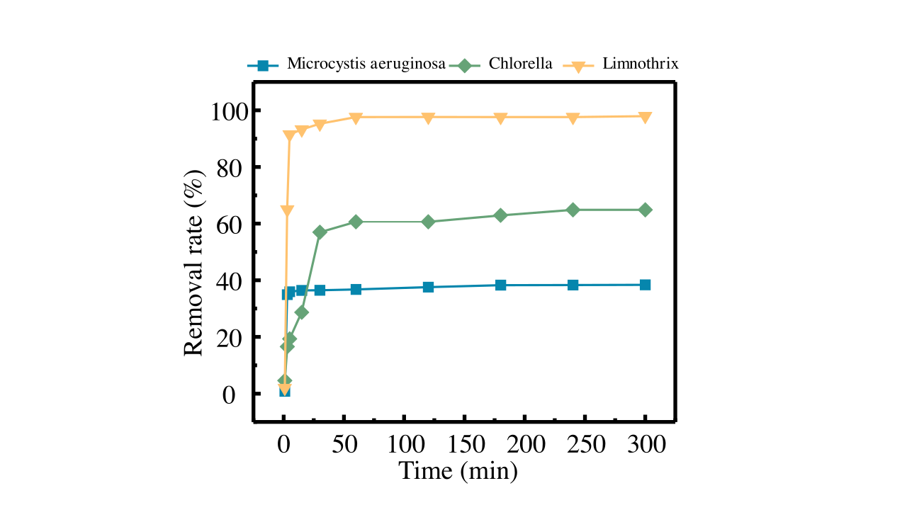
Supplementary Fig. 5
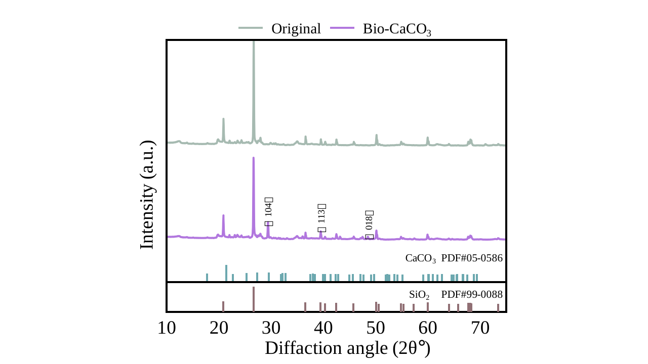
Supplementary Fig. 6
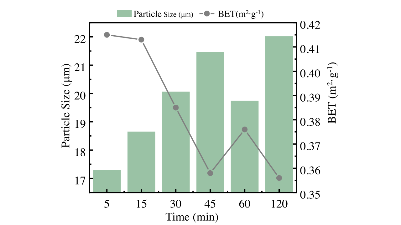
Supplementary Fig. 7
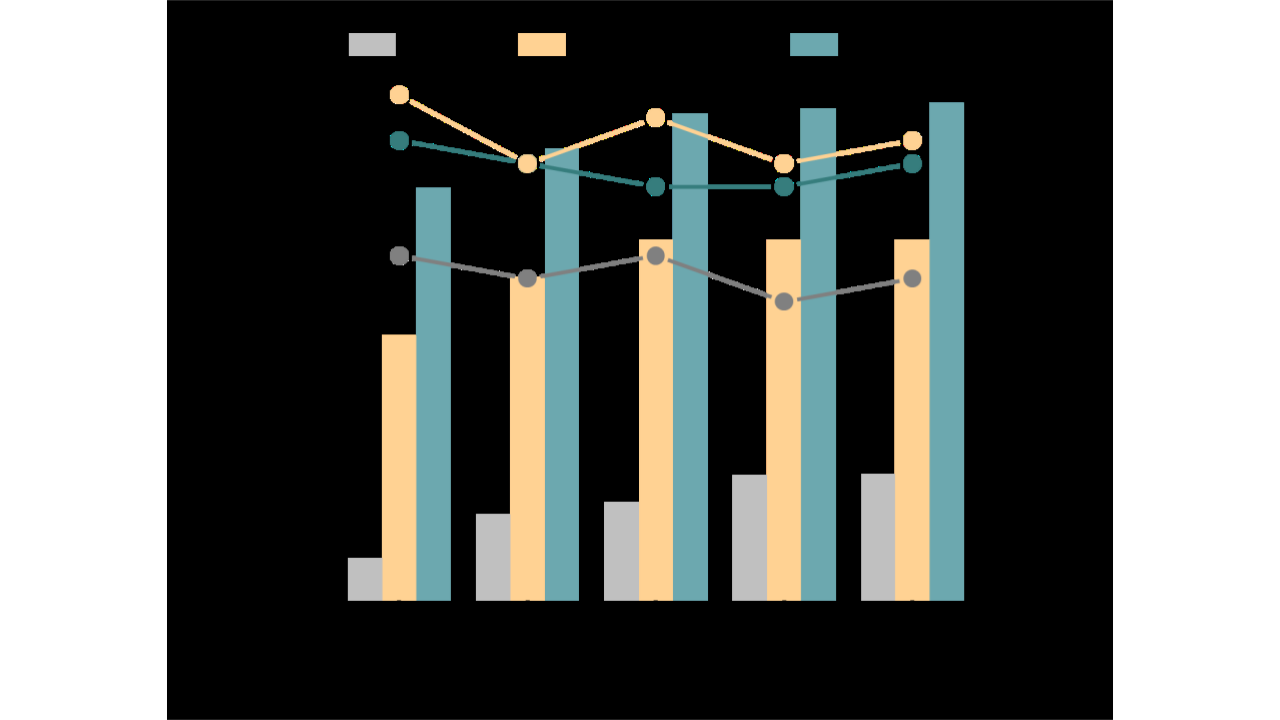
Supplementary Fig. 8
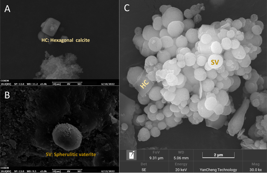
Supplementary Fig. 9
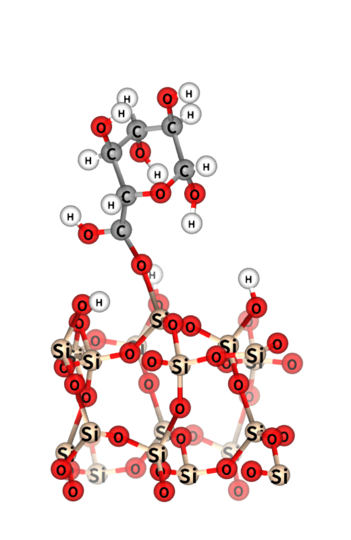
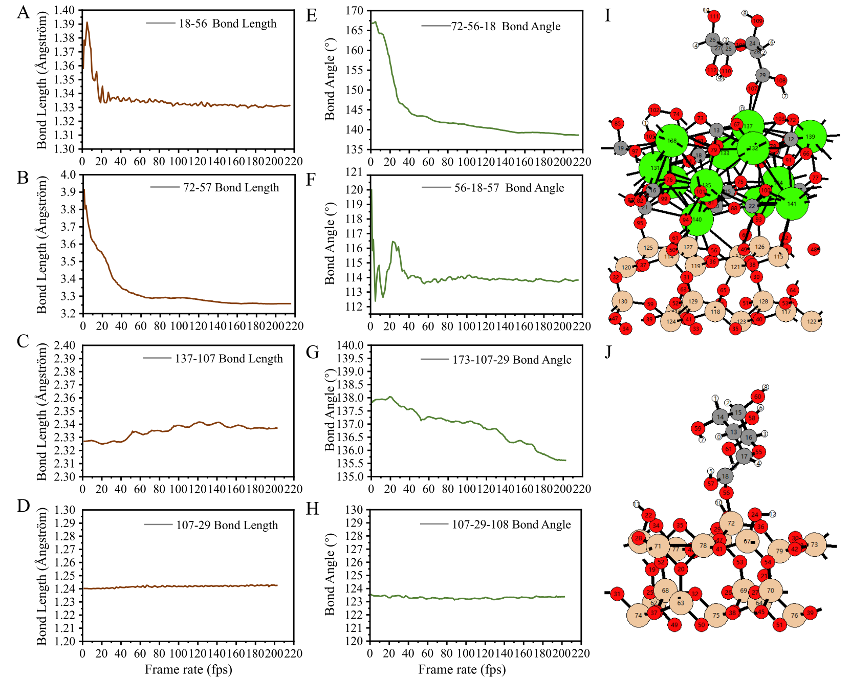
FigS9.gif). Time-resolved molecular dynamics snapshot showing the adsorption configuration of glucuronic acid on an unmodified silanol-rich silica surface. Silicon atoms are shown in beige, oxygen in red, carbon in grey, and hydrogen in white. The molecular conformation illustrates hydrogen bonding interactions between the hydroxyl/carboxyl groups of glucuronic acid and surface silanol groups, corresponding to the structural model used for bond length and bond angle trajectory analysis in Fig. S11A–D.
Supplementary Fig. 10
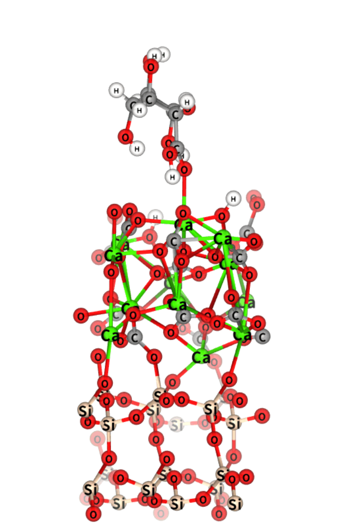
FigS10.gif). Atomic configuration from DFT molecular dynamics simulation showing the interaction between glucuronic acid and the bio-CaCO3–modified silica surface. Gray, red, white, beige, black, and green spheres represent C, O, H, Si, Ca–C carbonate groups, and Ca atoms, respectively. Ca atoms form coordination bonds with surface oxygen atoms and bridge carbonate groups to the silica substrate, illustrating the Ca–O–Si linkage and multi-point anchoring mechanism at the bio-CaCO3–modified interface. This structural model corresponds to that used for bond length and bond angle trajectory analyses shown in Fig. S11E–H.
Supplementary Fig. 11
Supplementary tables
Supplementary Table 1
| Ingredients | Content (%) | Unit |
|---|---|---|
| pH | 7.59±0.1 | — |
| TDS | 283.88±0.5 | mg L-1 |
| TH | 106.94±0.5 | mg L-1(CaCO3) |
| EC | 62.17±1.5 | μS cm-1 |
| TP | 20±0.3 | μg L-1 |
| TN | 3.2±0.04 | mg L-1 |
| COD(Mn) | 3.0±0.2 | mg L-1 |
| K+ | 0.91±0.03 | mg L-1 |
| Na+ | 9.55±0.05 | mg L-1 |
| Ca2+ | 83.53±0.2 | mg L-1 |
| Mg2+ | 25.96±0.1 | mg L-1 |
| Cl- | 12.99±0.03 | mg L-1 |
| SO42- | 12.27±0.02 | mg L-1 |
| HCO3- | 201.73±0.3 | mg L-1 |
Supplementary Table 2
| Ingredients | Content (%) |
|---|---|
| SiO2 | 68.72 |
| Al2O3 | 19.56 |
| Fe2O3 | 4.19 |
| MgO | 1.01 |
| CaO | 1.12 |
| Na2O | 0.75 |
| K2O | 2.9 |
| MnO | 0.06 |
| P2O5 | 0.22 |
| TiO2 | 0.86 |
| SO3 | 0.53 |
| Cl | 0.05 |
| Zn | 0.01 |
| Sr | 0.01 |
Supplementary Table 3
| Detection liquid | \(\gamma^{LW}\) | \(\gamma^{+}\) | \(\gamma^{-}\) | \(\gamma^{AB}\) | \(\gamma^{Tol}\) |
|---|---|---|---|---|---|
| Ultrapure water | 21.8 | 25.5 | 25.5 | 51.0 | 72.8 |
| Formamide | 39.0 | 2.28 | 39.6 | 19.0 | 58 |
| Diiodomethane | 50.8 | 0.0 | 0.0 | 0.0 | 50.8 |
Supplementary Table 4
| Factor | -1 | 0 | 1 |
|---|---|---|---|
| A: Added fraction of CaCO3 (%) | 2 | 8 | 14 |
| B: Residual free Ca2+ (%) | 0 | 50 | 100 |
| C: Time of reaction (min) | 10 | 60 | 110 |
Supplementary Table 5
| Time (min) | Chlorella vulgaris (μg L-1) | Microcystis aeruginosa (μg L-1) | Limnothrix sp. (μg L-1) | |||
|---|---|---|---|---|---|---|
| Control | Experimental | Control | Experimental | Control | Experimental | |
| 1 | 1139.4 | 1145.02 | 3245.2 | 3273.1 | 970.3 | 980.38 |
| 3 | 1142.7 | 1001.16 | 3252.6 | 2147.44 | 954.8 | 350.36 |
| 5 | 1135.3 | 968.22 | 3238.9 | 2113.0 | 940.1 | 85.24 |
| 15 | 1137.9 | 855.88 | 3248.7 | 2097.76 | 910.2 | 67.86 |
| 30 | 1140.1 | 516.42 | 3240.3 | 2095.94 | 885.4 | 47.4 |
| 60 | 1132.6 | 472.26 | 3235.8 | 2086.22 | 860.5 | 23.7 |
| 120 | 1139.0 | 472.26 | 3242.1 | 2059.28 | 880.2 | 23.54 |
| 180 | 1128.0 | 444.84 | 3238.0 | 2035.9 | 900.7 | 23.7 |
| 240 | 1133.8 | 421.3 | 3244.5 | 2035.58 | 920.9 | 23.7 |
| 300 | 1130.0 | 421.3 | 3239.9 | 2032.66 | 945.6 | 20.46 |
Supplementary Table 6
| Equilibrium parameters of adsorption | Adsorption | kinetics | ||||||
|---|---|---|---|---|---|---|---|---|
| Category | Langmuir | Freundlich | First-order kinetic | Secondary- orderkinetic | ||||
| KL | 1/n | KF | K1 | K2×10-6 | ||||
| (L μg-1) | (μg g−1) (L μg−1)1/n | (min-1) | (g (μg min)-1) | |||||
| Chlorella | Original | 0.009 | 0.418 | 40.378 | 0.014 | 6.35 | ||
| Bio- CaCO3 | 0.011 | 0.391 | 50.331 | 0.0184 | 6.05 | |||
| Microcystis aeruginosa | Original | 0.002 | 0.495 | 26.24 | 0.0168 | 2.689 | ||
| Bio- CaCO3 | 0.003 | 0.436 | 43.36 | 0.0185 | 2.280 | |||
| Limnothrix | Original | 0.004 | 0.841 | 12.24 | 0.0152 | 3.960 | ||
| Bio- CaCO3 | 0.003 | 0.964 | 13.29 | 0.0191 | 3.841 |
Supplementary Table 7
| Surface free energy parameters (mJ m-2) | Aii (×10-21 J) | |||||
|---|---|---|---|---|---|---|
| γ+ | γ- | γ | A132 (×10-21J) | Surface energy(mN m-1) | ||
| Modified Sediments | 0.6 | 31.61 | 39.99 | 57.58 | 5.41 | 48.69 |
| Algae organisms | 0.21 | 56.54 | 48.18 | 69.37 | 55.07 | |
| 5.12 | ||||||
| Unmodified Sediments | 0.08 | 34.72 | 38.88 | 55.99 | 42.21 | |
| MilliQ water | 25.5 | 25.5 | 21.8 | 31.39 |
Supplementary Table 8
| Surface/ Solution | Zeta potential | Contact angle (°) | Surface tension values ( mJ m-2) | ΔGAB | |||||
|---|---|---|---|---|---|---|---|---|---|
| (mV) | θW | θF | θD | γLW | γ+ | γ- | (mJ m-2) | ||
| Modified Sediments | -9.65 | 45°66’ | 34°62’ | 39°24’ | 39.99 | 0.60 | 31.61 | 31.282 | |
| Organisms | -5.78 | 7°2’ | 9°2’ | 18°6’ | 48.18 | 0.21 | 56.54 | ||
| 26.373 | |||||||||
| Unmodified Sediments | -14.6 | 49°46’ | 44°76’ | 42°26’ | 38.88 | 0.08 | 34.72 |
Supplementary Table 9
| Factor 1 | Factor 2 | Factor 3 | Response 1 | Response 2 | ||
|---|---|---|---|---|---|---|
| Std | Run | Mass fraction of CaCO3 | Residual free Ca2+ (%) | Time | Y1: Chl-a removal rate | Y2: COD removal rate |
| (%) | - | (min) | (%) | (%) | ||
| 1 | 12 | 2 | 0 | 60 | 73.76 | 73.99 |
| 2 | 8 | 14 | 0 | 60 | 87.58 | 68.21 |
| 3 | 2 | 2 | 100 | 60 | 76.59 | 84.6 |
| 4 | 16 | 14 | 100 | 60 | 90.25 | 42.77 |
| 5 | 6 | 2 | 50 | 10 | 75.31 | 79.77 |
| 6 | 9 | 14 | 50 | 10 | 84.59 | 65.9 |
| 7 | 7 | 2 | 50 | 110 | 80.05 | 90.17 |
| 8 | 13 | 14 | 50 | 110 | 93.47 | 71.68 |
| 9 | 17 | 8 | 0 | 10 | 82.74 | 79.77 |
| 10 | 10 | 8 | 100 | 10 | 85.23 | 50.94 |
| 11 | 4 | 8 | 0 | 110 | 87.29 | 63.58 |
| 12 | 11 | 8 | 100 | 110 | 92.3 | 76.3 |
| 13 | 14 | 8 | 50 | 60 | 92.89 | 87.98 |
| 14 | 1 | 8 | 50 | 60 | 93.78 | 84.89 |
| 15 | 5 | 8 | 50 | 60 | 92.94 | 90.88 |
| 16 | 3 | 8 | 50 | 60 | 93.06 | 86.12 |
| 17 | 15 | 8 | 50 | 60 | 93.83 | 90.32 |
Supplementary Table 10
| Mass fraction of CaCO3(%) | Residual free Ca2+(%) | Time of reaction(min) | Removal- Chl-a (%) | Removel-COD (%) | |||
|---|---|---|---|---|---|---|---|
| Observed | Predicted | Observed | Predicted | ||||
| 7.51 | 56 | 85.35 | 92.67 | 93.83 | 87.23 | 88.64 | |
| 7.51 | 56 | 85.35 | 91.98 | 93.83 | 86.54 | 88.64 |
References
Using bio-based CaCO3 functionalized sediment to simultaneously remove algae and COD through adsorption and sedimentation in water source reservoirs – du2025using: PUB Using bio-based CaCO3 functionalized sediment to simultaneously remove algae and COD through adsorption and sedimentation in water source reservoirs – du2025using: PUB Using bio-based CaCO3 functionalized sediment to simultaneously remove algae and COD through adsorption and sedimentation in water source reservoirs – du2025using: PUB du2025using: PUB Supplementary Information Supplementary Information
Almomani, F., Bhosale, R.R., 2021. Bio-sorption of toxic metals from industrial wastewater by algae strains Spirulina platensis and Chlorella vulgaris: Application of isotherm, kinetic models and process optimization. Science of The Total Environment 755, 142654. https://doi.org/10.1016/j.scitotenv.2020.142654
Anderson-Cook, C.M., Borror, C.M., Montgomery, D.C., 2009. Response surface design evaluation and comparison. Journal of Statistical Planning and Inference 139, 629–641. https://doi.org/10.1016/j.jspi.2008.04.004
Grimme, S., Antony, J., Ehrlich, S., Krieg, H., 2010. A consistent and accurateab initioparametrization of density functional dispersion correction (DFT-d) for the 94 elements h-pu. The Journal of Chemical Physics 132. https://doi.org/10.1063/1.3382344
Huang, Y., Wang, Y., Huang, Y., Zhang, L., Ye, F., Wang, J., Shang, J., Liao, Q., 2020. Impact of sediment characteristics on adsorption behavior of typical antibiotics in lake taihu, china. Science of The Total Environment 718, 137329. https://doi.org/10.1016/j.scitotenv.2020.137329
Kresse, G., Furthmüller, J., 1996. Efficient iterative schemes forab initiototal-energy calculations using a plane-wave basis set. Physical Review B 54, 11169–11186. https://doi.org/10.1103/physrevb.54.11169
Monkhorst, H.J., Pack, J.D., 1976. Special points for brillouin-zone integrations. Physical Review B 13, 5188–5192. https://doi.org/10.1103/physrevb.13.5188
Novak, J.T., Brune, D.E., 1985. Inorganic carbon limited growth kinetics of some freshwater algae. Water Research 19, 215–225. https://doi.org/10.1016/0043-1354(85)90203-9
Oss, C.J. van, 1995. Hydrophobicity of biosurfaces — origin, quantitative determination and interaction energies. Colloids and Surfaces B: Biointerfaces 5, 91–110. https://doi.org/10.1016/0927-7765(95)01217-7
Oss, C.J. van, 1993. Acid—base interfacial interactions in aqueous media. Colloids and Surfaces A: Physicochemical and Engineering Aspects 78, 1–49. https://doi.org/10.1016/0927-7757(93)80308-2
Perdew, J.P., Burke, K., Ernzerhof, M., 1996. Generalized gradient approximation made simple. Physical Review Letters 77, 3865–3868. https://doi.org/10.1103/physrevlett.77.3865
Veith, J.A., Sposito, G., 1977. On the use of the langmuir equation in the interpretation of “adsorption” phenomena. Soil Science Society of America Journal 41, 697–702. https://doi.org/10.2136/sssaj1977.03615995004100040015x